Author Ni Yang, PhD. JH Technologies, Lab Scientist
If you’ve ever wandered around Fremont, California, sniffling with tissues in hand and allergy meds in your pocket, you already know that pollen is the invisible foe of spring and summer. But have you ever paused mid-sneeze—eyes watering, nose running—and wondered: What does this tiny troublemaker actually look like up close?
Well, hold onto your microscope slides, because what we found was equal parts bizarre, beautiful, and unexpectedly fascinating.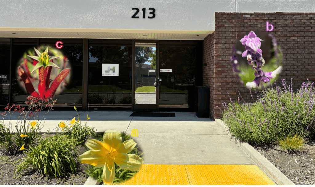
First Look: Peeking at Pollen Under the Optical Microscope
We started our investigation just outside the JH Technology office—because why go far when science begins right at your doorstep?
Using our Leica DM8000 M optical microscope, we examined flowers growing around the office, including:
- Yellow Cocotte Lily
- Mexican Bush Sage (Salvia leucantha)
- Red Kangaroo Paw (Anigozanthos)
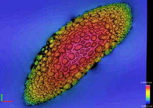
Yellow Cocotte Lily pollen appeared large, oval, and golden—almost glowing. Its textured surface gave it a grainy sparkle. Figure (a)
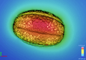
Mexican Bush Sage showed smaller, rounder grains with a fuzzy, granular look, matching its delicate petals. Figure (b)
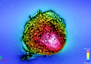
Red Kangaroo Paw, on the other hand, lived up to its exotic name—its pollen looked spiky and irregular, like tiny golden starbursts. Figure (c)

Already, we were seeing just how much variety lives in the pollen world—but things only got more interesting from here.
Zooming In: High-Resolution Detail with Scanning Electron Microscopy (SEM)
Next, we cranked up the detail using our CIQTEK FE-SEM5000X field emision scanning electron microscope. SEM imaging reveals micro and nanoscale surface textures invisible to both the naked eye and optical microscopes. Because pollen is organic and non-conductive, we coated it with a thin 4 nm layer of gold–palladium (80/20) using the Leica EM ACE600 sputter coater to enable effective imaging in the CIQTEK SEM5000X.


CIQTEK FE SEM 5000X SEM Leica EM ACE600

What We Saw Under SEM:
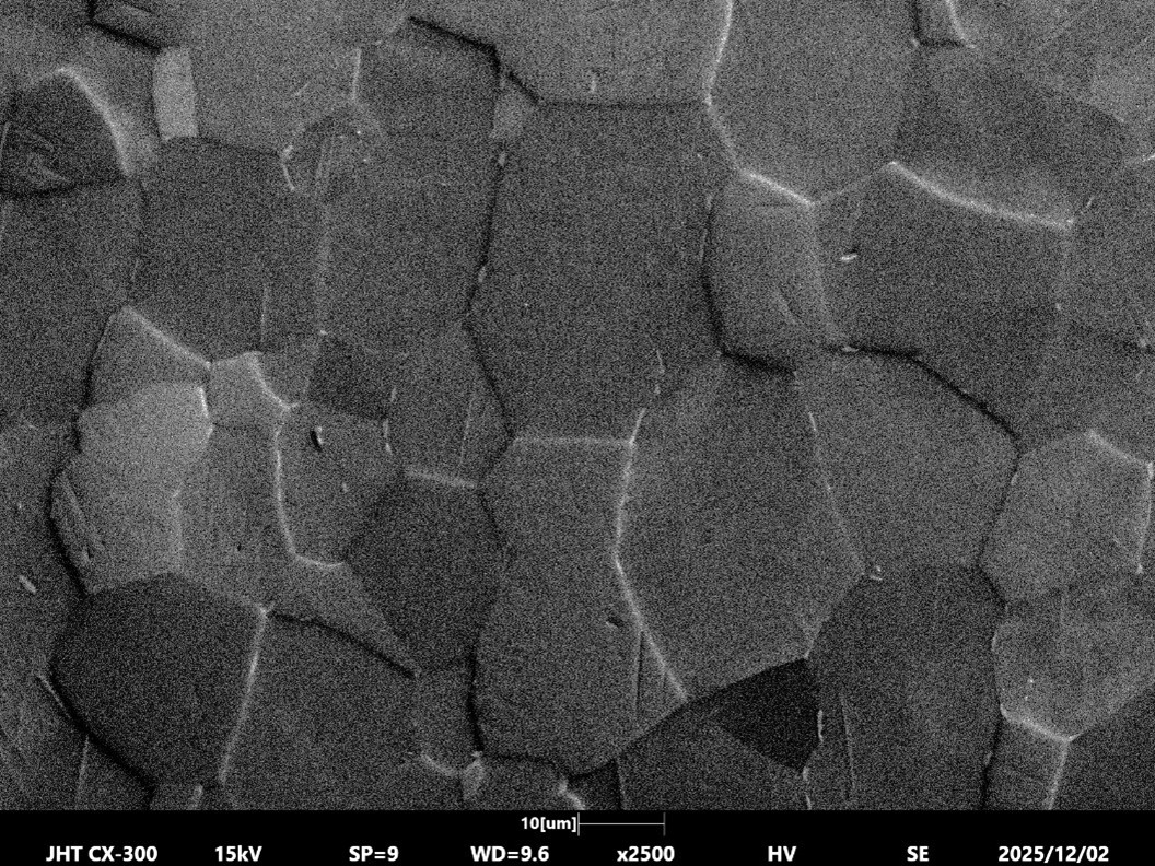
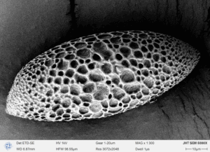
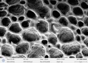
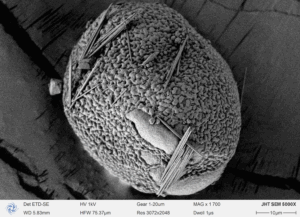
Yellow Lily Pollen
- Size: ~40 µm × 100–110 µm
- Surface: Reticulate (net-like) pattern with polygonal pits (1–10 µm wide) and blister-like protrusions on the surface.
- Visual vibe: Walls between these pits are relatively thick, resembling an irregular honeycomb-like, bumpy appearance.
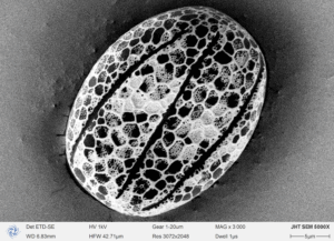
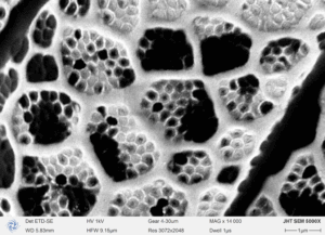
Mexican Bush Sage Pollen
- Shape: Prolate (think watermelon-shaped)
- Features: Net-like exine pattern surface, plus three long furrows—creating a “honeycomb-within-a-honeycomb” morphology.
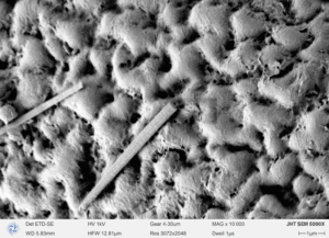
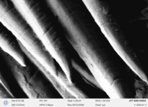
Red Kangaroo Paw Pollen
- Shape: Mostly spherical to slightly oval
- Surface: Wrinkled, rough, with needle-like structures likely made of calcium oxalate crystals (raphides) [1].
- The fine ridges and parallel alignment of these structures give it a distinct, almost armored appearance.
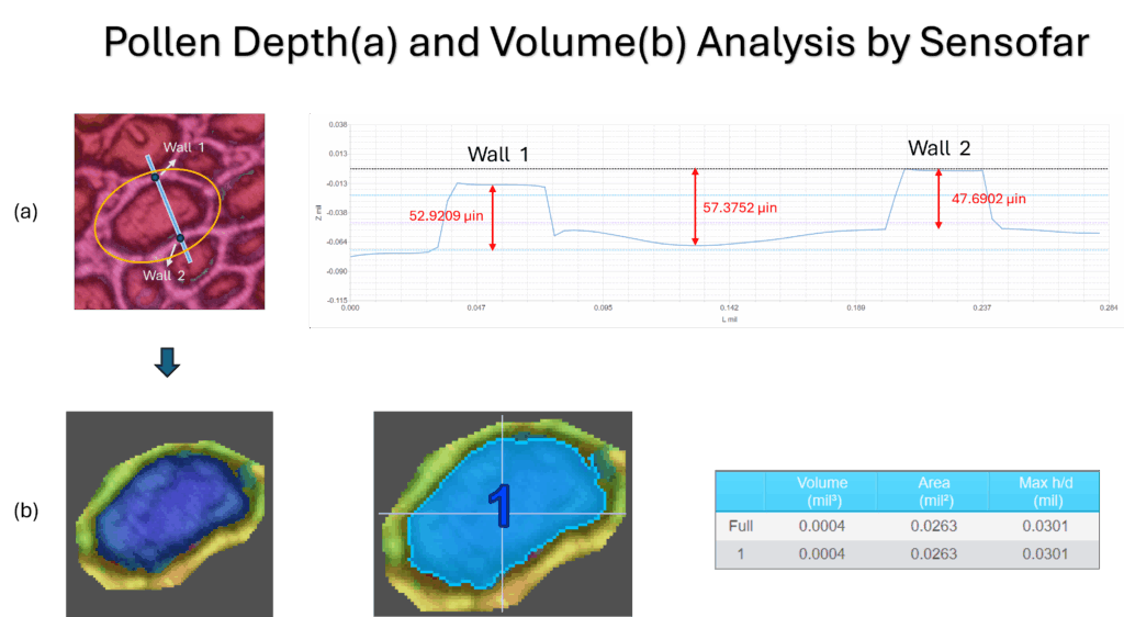
Bringing in 3D: Confocal Imaging with Sensofar
While SEM gives stunning surface detail, it doesn’t provide quantative data such volume, depth, or true-scale 3D topography. That’s where the Sensofar S neox came in.
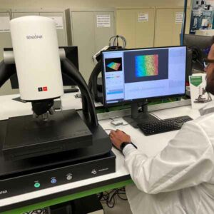 Sensofar-sneox
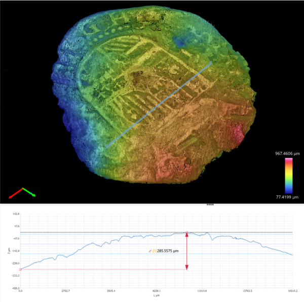
This advanced system creates 3D surface height maps with vertical resolution down to 0.16 µm, giving us quantifiable data like:
- Mesh depth and wall height
- Surface roughness
- Volume of pits or textures
Yellow Cocotte Lily Mexican Bush Sage
Red Kangaroo Paw Pollen
Results:
- The confocal images mirrored SEM findings, but with added color-coded depth maps.
- Using the Yellow Lily as an example, we could precisely measure the depths of surface walls and mesh, as well as the mesh volumes.
In short: SEM shows you what’s there; the 3D confocal sytem tells you how much and how deep.
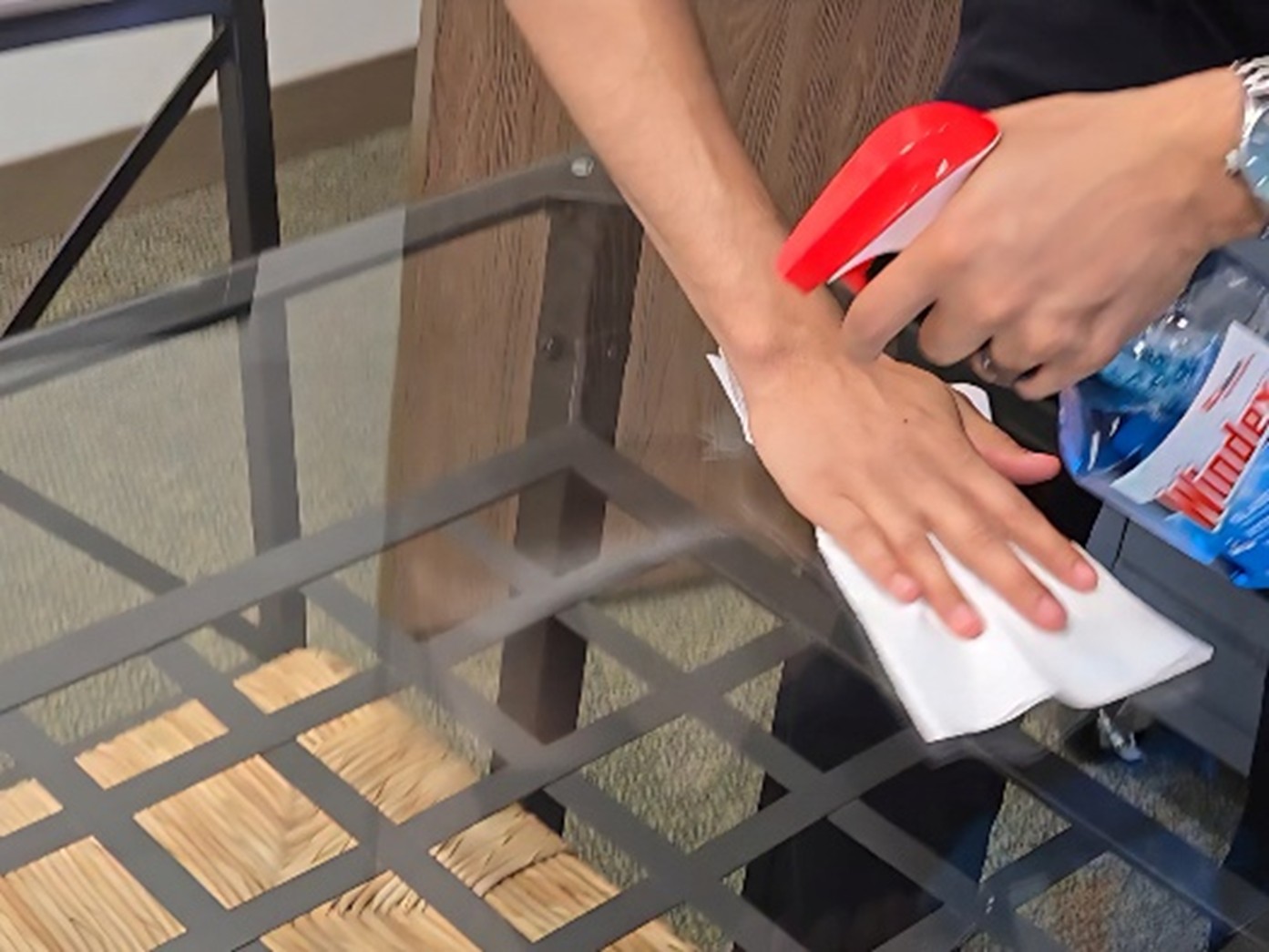
Bonus Round: Pollen on Our Company Car
Curious about what airborne pollen might be hitching a ride on our vehicles, we scraped a thin sample off the front windshield of the JH company car.
What We Found:
- A chaotic mix of dust, debris, and pollen—like a microscopic farmer’s market.
- Among the clutter, we discovered mystery pollen grains, likely from local grasses or weeds.
- SEM revealed:
- Spherical and oval grains
- Intricate textures: ridges, grooves, and porous surfaces at the submicron level
Turns out, our car was a tiny ecosystem on wheels.
Time for a car wash!

Conclusion: The Hidden World Beneath Your Nose
This journey into Fremont’s pollen landscape revealed just how complex and diverse the microscopic world really is.
With help from Leica optical microscopy, CIQTEK SEM, and Sensofar confocal analysis, we were able to:
- Visualize pollen in stunning detail
- Quantify surface structure and depth
- Discover unexpected pollen types hitching rides in everyday places
So next time you’re battling allergies, remember—those sneeze-inducing particles are also tiny works of art, each with its own story, structure, and microscopic charm.
References
[1] https://en.wikipedia.org/wiki/Raphide