Author Ken Hirsch, Yi Zhang (PhD), JH Technologies, Applications Scientists
Faster Analysis with Less Sample Preparation: A Practical Summary
 Paper Towel No Coat Low Vac
EXECUTIVE SUMMARY
For decades, Scanning Electron Microscopy (SEM) has been one of the most powerful techniques for analyzing materials at the microscopic level. Whether examining polymers, biological samples, coatings, or industrial components, SEM provides detailed insights into surface structure, morphology, and composition.
However, traditional SEM imaging often comes with a challenge: many samples require extensive preparation before they can be analyzed.
The Problem with Non-Conductive Samples
Materials such as plastics, paper, biological tissues, ceramics, and coatings are electrically insulating. In a conventional high-vacuum SEM, these materials can accumulate charge during imaging, causing distorted images, poor contrast, and unreliable results.
To overcome this, operators often apply conductive coatings such as gold or carbon. While effective, these preparation steps add time, cost, specialized equipment requirements, and may alter the sample itself.
A Simpler Alternative: Low Vacuum SEM
Results
A recent study using the Coxem EM-40 Desktop SEM compared traditional high-vacuum imaging with Low Vacuum imaging across several common sample types:
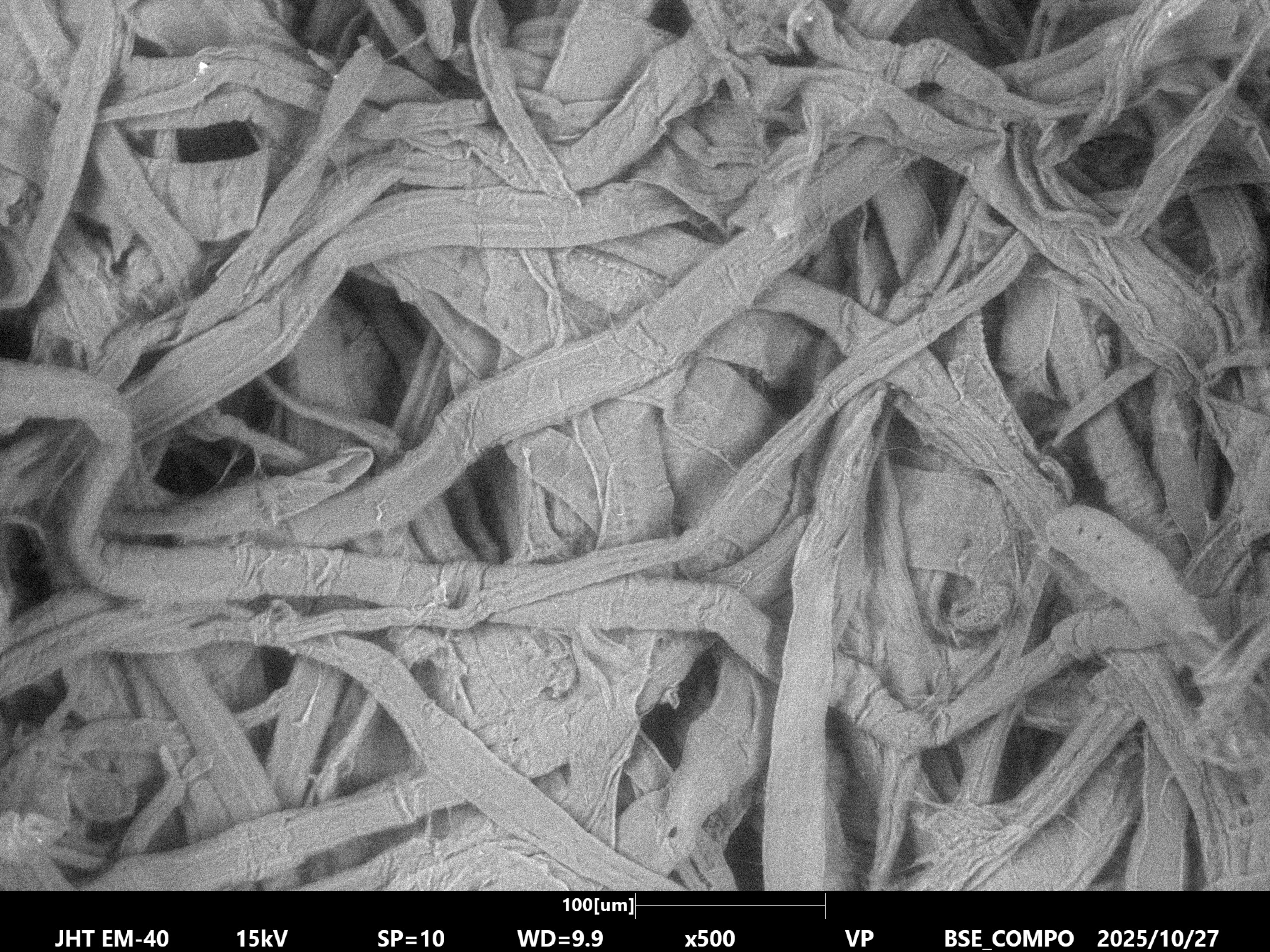
Case Study 1: Paper Fibers
Paper Towel: No Coat, Low Vac
Case Study 2: Human Hair (Beam-Sensitive Biological Material)
LV mode reduced both charging and beam-induced damage on the cuticle structure, making it the only viable option for examining uncoated hair or similar delicate biological materials (see comparison images in the full white paper).
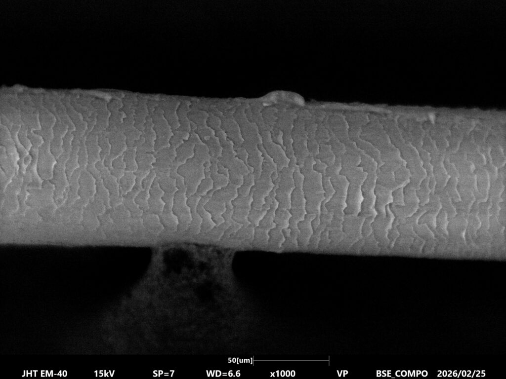 Human Hair: No Coat, Low Vac
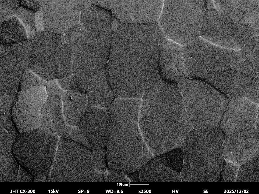
Case Study 3: Diatom Algae (Fine Biological Microstructure)
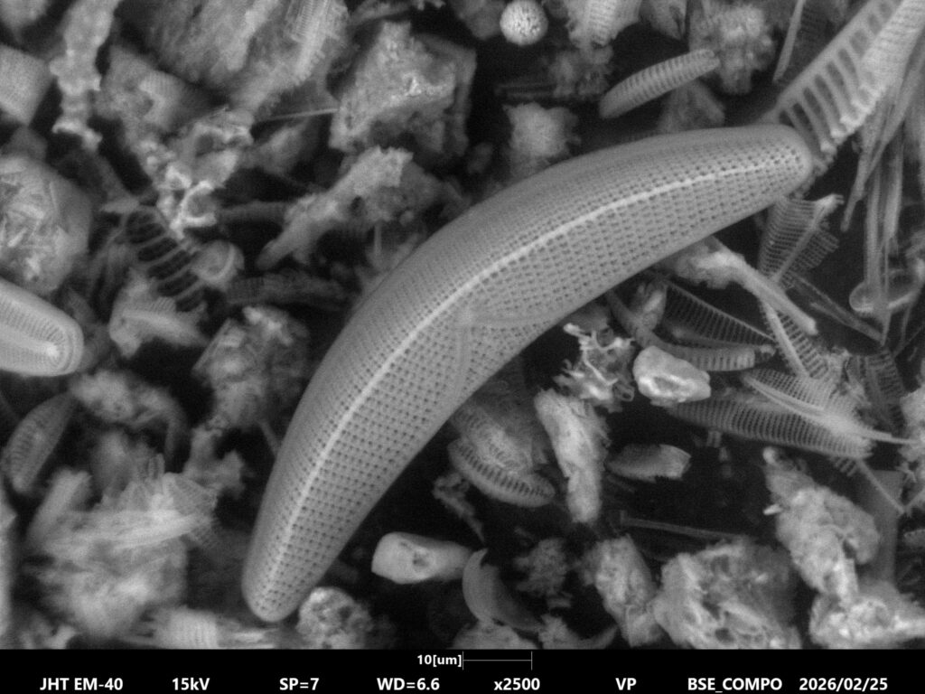 Diatom: No Coat, Low Vac
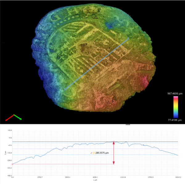
Case Study 4: Epoxy-Mounted Paint Cross Section (Layered Industrial Material)
LV mode enabled fast cross-sectional analysis without coating, ideal for quality control situations where turnaround time matters (see comparison images in the full white paper).
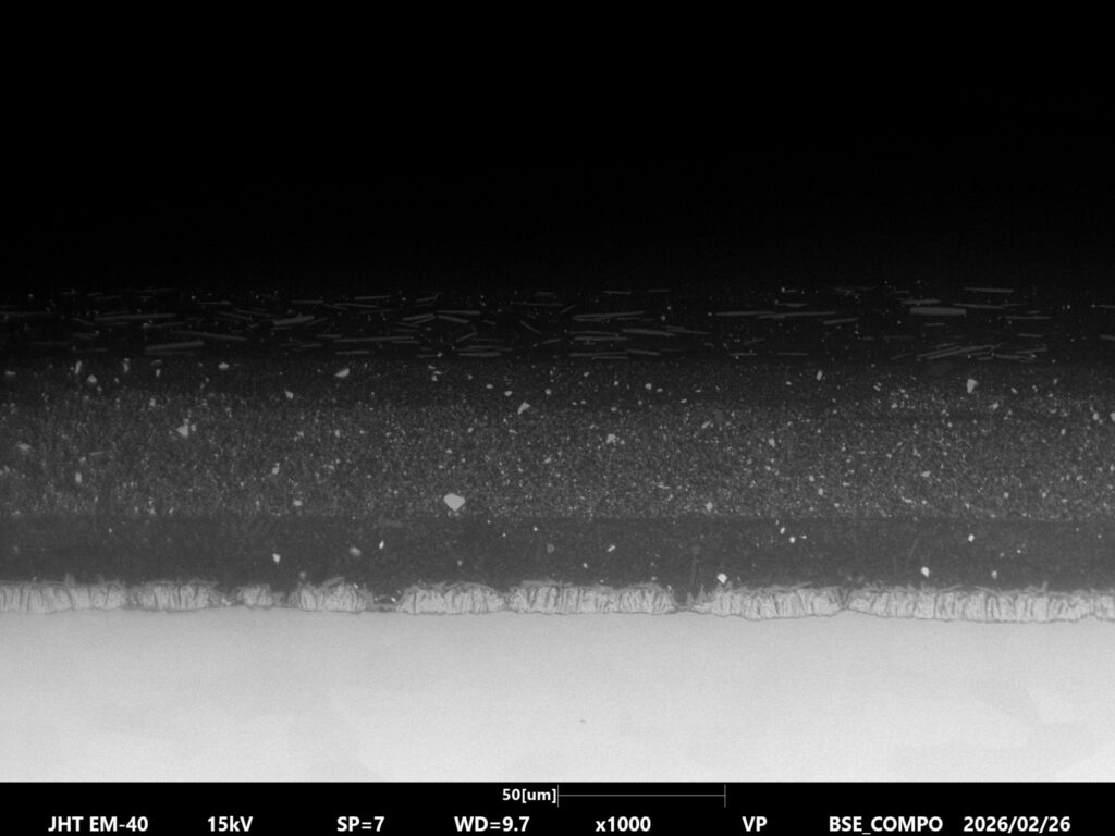 Car Paint: No Coat, Low Vac
When Should You Use Low Vacuum SEM?
Low Vacuum mode is particularly valuable when:
- Samples are non-conductive
- Materials are delicate or beam-sensitive
- Fast turnaround is important
- Samples must remain unaltered for additional testing
Traditional high-vacuum imaging remains the preferred option when maximum resolution is the primary objective.
For many routine applications, however, Low Vacuum SEM delivers more than enough resolution while significantly simplifying the workflow.